1. Home
  2. Procedures
  3. Retina, Uvea & Vitreous

Retina, Uvea & Vitreous

Caring for the back of your eye.

Caring for the back of your eye

The back segment of the eye

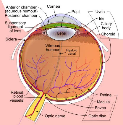
The retina, uvea, and vitreous form the back segment of the eye — the delicate structures responsible for capturing and processing light into the images we see.

The retina is the light-sensitive layer that converts light into neural signals, the uvea is the middle, blood-vessel-rich layer that nourishes the eye, and the vitreous is the clear gel filling the eye’s interior.

Conditions affecting these structures can develop silently and, without timely treatment, may lead to permanent vision loss — making early detection and specialised care essential.

Labelled cross-section of the human eye showing the retina, macula, uvea (iris, ciliary body, choroid), vitreous humour and optic nerve
Structures of the eye: the retina, uvea and vitreous make up the back segment.

Conditions we manage

Conditions we manage

Diabetic retinopathy

A common complication of diabetes where high blood sugar damages the tiny blood vessels in the retina, causing them to leak, swell, or grow abnormally. It progresses in stages — from mild non-proliferative changes to severe proliferative disease — and is a leading cause of vision loss in working-age adults.

Age-related macular degeneration (AMD)

A progressive condition affecting the macula, the part of the retina responsible for sharp, central vision. AMD occurs in two forms — dry AMD (gradual thinning of retinal tissue) and wet AMD (abnormal blood vessel growth that leaks fluid) — and is a leading cause of vision loss in people over 60.

Emergency

Retinal detachment

A serious, sight-threatening emergency where the retina separates from the underlying tissue that supports it. Warning signs include sudden flashes of light, a shower of new floaters, or a shadow or curtain moving across the field of vision — this requires immediate treatment.

Uveitis

Inflammation of the uvea, which can be caused by infection, autoimmune disease, or injury. Symptoms include eye pain, redness, blurred vision, light sensitivity, and floaters. Left untreated, uveitis can lead to complications like glaucoma, cataracts, or permanent vision loss.

Urgent

Retinal vein & artery occlusion

Blockages in the blood vessels supplying the retina, which can cause sudden, painless vision loss and require prompt evaluation and management.

Macular hole & epiretinal membrane

Structural changes to the macula — a hole or a wrinkling membrane — that can distort or blur central vision, often requiring surgical repair.

Floaters and vitreous disorders

Age-related changes or shrinkage of the vitreous gel can cause floaters and flashes; while often harmless, a sudden increase warrants prompt evaluation to rule out retinal tears.

Signs to watch for

Symptoms that need attention

  • Sudden flashes of light or a sudden increase in floaters
  • A shadow or curtain effect in your vision
  • Blurred, distorted, or wavy central vision (straight lines appearing bent)
  • Difficulty seeing in low light or a blind spot in central vision
  • Eye pain, redness, or light sensitivity
  • Sudden, painless loss of vision in part or all of the visual field

Diagnosis

Diagnostic technology

Optical Coherence Tomography (OCT)
High-resolution cross-sectional imaging of the retina to detect fluid, swelling, or structural changes
Fundus Fluorescein Angiography (FFA)
Highlights blood flow and leakage in retinal vessels
Fundus photography
Detailed imaging to document and monitor retinal health over time
B-scan ultrasound
Used when the view of the retina is obscured, such as by bleeding
Visual field testing
Assesses functional vision loss

Treatment

Treatment approaches

ConditionTreatment options
Diabetic retinopathyBlood sugar control, anti-VEGF injections, laser photocoagulation, vitrectomy in advanced cases
Wet AMDAnti-VEGF injections to stop abnormal vessel growth and leakage
Dry AMDNutritional supplements (AREDS formula), lifestyle modification, close monitoring
Retinal detachmentEmergency surgery — laser, cryotherapy, pneumatic retinopexy, vitrectomy, or scleral buckle
UveitisAnti-inflammatory drops or injections, immunosuppressive therapy for underlying causes
Macular hole / membraneVitrectomy surgery to repair and restore central vision

What to expect

Many retinal conditions require ongoing monitoring even after treatment begins, since conditions like diabetic retinopathy and wet AMD are chronic and managed over time rather than cured in a single visit. Treatment plans are tailored to the specific condition, its severity, and how it’s affecting your vision, often involving a combination of injections, laser therapy, and lifestyle management.

Why early detection matters

Many retinal and uveal conditions progress silently until significant vision loss has occurred. Regular dilated eye exams — especially for people with diabetes, high blood pressure, or a family history of AMD — allow early detection when treatment is most effective at preserving vision.

Your care at Kunj Eye Hospital

Doctors, equipment & related services

Your doctors

Consultations are with our ophthalmologists:

Equipment used

OCT scans, fundus photography, fundus fluorescein angiography, indirect ophthalmoscope, B-scan ultrasound and PRP laser.

See all our facilities & equipment

Ready to take the first step towards clearer, healthier vision with expert care?